MULTI-SUBSTRATE ENZYMES
In reality, many enzymes have more than one substrate (A, B) and more than one product (P, Q). For
example, the enzyme alcohol dehydrogenase catalyzes the oxidation of ethanol with NAD (a biological oxidizing agent) to form
acetaldehyde and NADH. How do you do enzymes kinetics on these more complicated systems? The answer is fairly
straightforward. You keep one of the substrates (B) fixed, and vary the other substrate (A) and obtain a series
of hyperbolic plots of vo vs A at different fixed B concentrations. This would give a series of linear 1/v
vs 1/A double-reciprocal plots (Lineweaver-Burk plots) as well. The pattern of Lineweaver-Burk plots depends on how
the reactants and products interact with the enzyme.
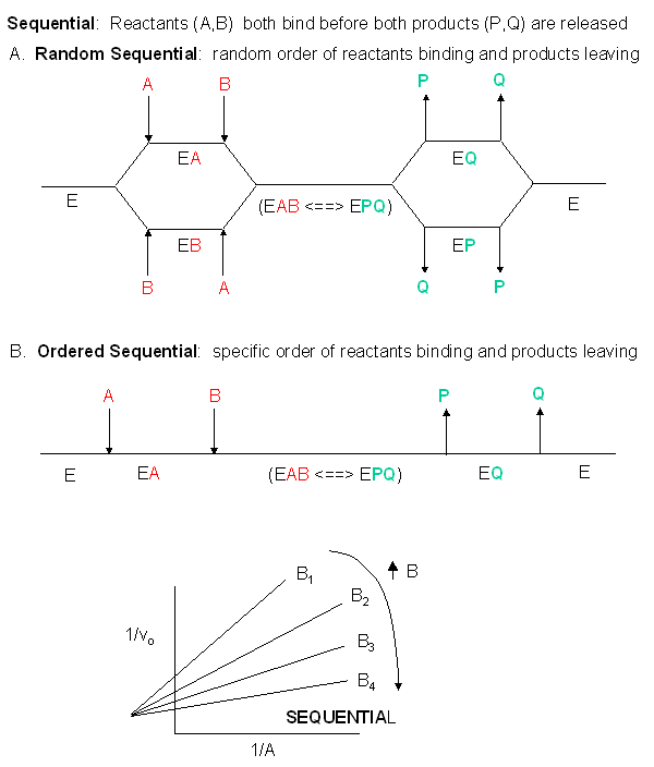
Sequential Mechanism: In this mechanism, both substrates must bind to
the enzyme before any products are made and released. The substrates might bind to the enzyme in a random
fashion (A first then B or vice-versa) or in an ordered fashion (A first followed by B). An abbreviated
notation scheme is shown below for the sequential random and sequential ordered mechanisms.
For both mechanisms, Lineweaver-Burk plots at varying A and different fixed values of B give a series of intersecting
lines.

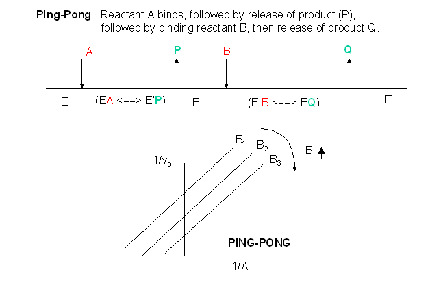
Ping-Pong Mechanism: In this mechanism, one substrate bind first to the enzyme
followed by product P release. Typically, product P is a fragment of the original substrate A. The rest of the
substrate is covalently attached to the enzyme E , which we now designate as E'. Now the second reactant, B, binds
and reacts with the enzyme to form a covalent adduct with the covalent fragment of A still attached to the enzyme to form
product Q. This is now released and the enzyme is restored to its initial form, E. This mechanism is term ping-pong.
An abbreviated notation scheme is shown below for the ping-pong mechanisms. For this mechanisms,
Lineweaver-Burk plots at varying A and different fixed values of B give a series of parallel lines. (An example of this
type might be human adipoctye acid phosphate, in which A, p-nitrophenylphosphate, binds to the enzyme covalently with the
expulsion of the product P, the p-nitrophenol leaving group. Water (B) then comes in and covalently attacks the enzyme,
forming an adduct with the phosphate which is covalently bound to the enzyme, releasing it as inorganic phosphate. In
this particular example, however, you can't vary the water concentration and it would be impossible to generate the parallel
Lineweaver-Burk plots characteristic of ping-pong kinetics.

ALLOSTERIC ENZYMES
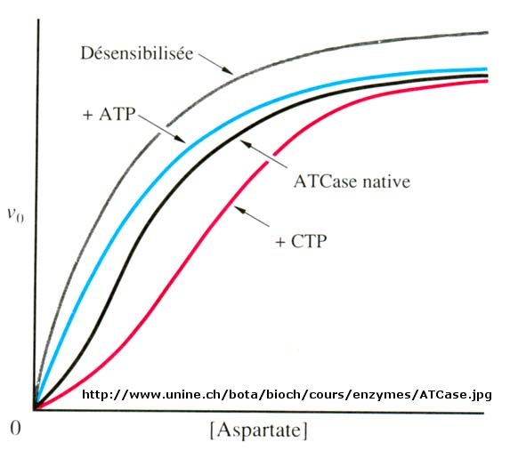
Many enzymes do not demonstrate hyperbolic saturation kinetics, or typical Michaelis-Menten kinetics.
Graphs of initial velocity vs substrate demonstrate sigmoidal dependency of v on S, much as we discussed with hemoglobin binding
of dioxygen. Enzymes that display this non Michaelis-Menten behavior have common characteristics. They :
- are multi-subunit
- bind other ligands at sites other than the active site (allosteric sites)
- can be either activated or inhibited by allosteric ligands
- exist in two major conformational states, R and T
- often control key reactions in major pathways, which must be regulated.
Examples of these enzymes include glycogen phosphorylase (the enzyme which breaks down intracellular glycogen
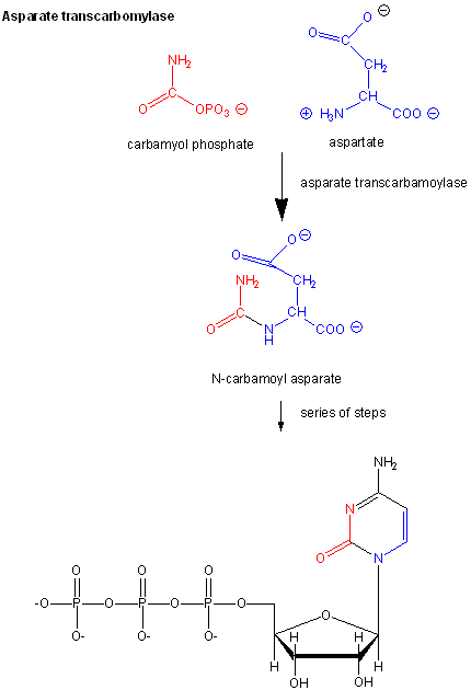
reserves) and aspartate transcarbamyolase, which catalyzes the first step in the synthesis of pyrimidine nucleotides. CTP
is an allosteric inhibitor of this enzyme, which makes physiological sense since high levels of this pyrimidine nucleotide
should inhibit the first enzyme in the synthesis of pyrimidines. ATP is a allosteric activator. This also makes sense since
if high levels of the purine nucleotide ATP are present, one would also want to balance the level of pyrimidine nucleotides.
Figure: Aspartate transcarbamoylase: reactions

Figure: Aspartate transcarbamoylase: Non Michaelis-Menten Kinetics

Earlier we saw that cooperative binding equilibrium could be modeled with the Hill Equation,
which we introduced through the equation Y = Ln/(Kd + Ln) = Ln/(P50n
+ Ln) where n is the cooperativity or Hill Coefficient. Likewise, for an enzyme which demonstrates cooperative
(sigmoidal) initial rates plots,
vo = VmSn/K0.50n + Ln)
When n=1, the equation reduces to the classical hyperbolic Michaelis Menten equation. For values
of n>1, sigmoidal plots are observed. We found a more easily understandable molecular interpretation of cooperative
binding of oxygen to hemoglobin using the MWC model (T and R states). The MWC model has also been applied successfully
to multi-subunit enzymes which display cooperative, sigmoidal kinetics. In this model, allosteric inhibitors (which
often don't resemble the substrate) bind preferentially to the T state, leading to lower activity, while allosteric
activators bind preferentially to the R state, leading to greater activity. Activators shift the vo vs S
curve to the left while inhibitors shift it to the right (much like protons and carbon dioxide in hemoglobin binding).
These allosteric ligands induce their effects by shifting the To <=> Ro equilibrium.
How do allosteric effectors change Vm and Km?
We have just studied how competitive, uncompetitive, and noncompetitive (or mixed) inhibitors influence
the apparent Km and Vm values for enzymes that display Michaelis-Menten kinetics. How are Vm and Km influenced in allosteric
enzymes? The examples given above, analogous to effects observed in hemoglobin:oxgyen binding, influence the apparent
Km, but not the Vm. (Go back to the hemoglobin binding curves and note that they shifted to the left or right, but all
reached a plateau at the same fractional saturation value of 1. Allosteric enzyme systems that behave like this are
called K systems. Allosteric enzymes in which effectors change Vm, called V systems, are also known.
V systems display hyperbolic vo vs S curves in which activators display a greater apparent Vm and
inhibitors display a lower Vm without affecting the apparent Km. In these systems, both the T and R forms have the same
affinity for substrate (hence the same apparent Km) but display different catalytic rate constants, kcat, for turnover
of the bound substrate (hence different apparent Vm values). In addition, the activator A and inhibitor I bind to the
T and R forms with different affinity, which again shifts the To <=> Ro equilibrium in the presence
of the allosteric effectors.
Cooperative binding of dioxygen to hemoglobin, regulated by allosteric effectors (protons and carbon dioxide),
was ideal for an oxygen transport system which must load and unload oxygen over a narrow range of oxygen concentrations and
allosteric effectors. Allosteric enzymes are usually positioned at key metabolic steps which can be regulated to activate
or inhibit whole pathways.
Web Links
Enzyme Regulation by Covalent Modification
Many enzymes are regulated not by allosteric ligands (activators and inhibitors), but by covalent modification.
Often the covalent modification involves phosphorylation (by enzymes called kinases which transfer a phosphate from
ATP to a Ser, Thr, or Tyr on the target enzyme) or phosphatases (which remove the phosphates from phospho-Ser, Thr,
or Tyr in the target protein). In fact 1-2% of all genes in the human genome code for kinases and phosphatases.

