|
|

adenine

thymine

guanine

cytosine
| |
|

DNA is the basic hereditary material
in all cells and contains all the information necessary to make proteins.
DNA is a linear polymer that is made up of nucleotide units . The nucleotide unit consists of a base, a
deoxyribose sugar, and a phosphate. There are four types of bases: adenine (A), thymine (T), guanine (G), and cytosine (C).
Each base is connected to a sugar via a ß glycosyl linkage. The nucleotide units are connected via the O3' and O5' atoms forming
phosphodiester linkages.
In normal DNA, the bases form pairs: A to T and G to C. This is called complementarity. A duplex of DNA
is formed by two complementary chains that are arranged in an anti-parallel manner.
The results of fiber and single crystal x-ray crystallographic studies have shown that DNA can have several
conformations. The most common one is calledB-DNA. B-DNA is a right-handed double helix with a
wide and narrow groove. The bases are perpendicular to the helix axis.
DNA can also be found in theA form in which the major groove is very deep and
the minor groove is quite shallow.
A very unusual form of DNA is the left-handed Z-DNA. In this DNA, the basic
building block consists of two nucleotides, each with different conformations. Z-DNA forms excellent crystals.
Several years ago it was discovered that nucleic acids can form four stranded structures
and a few examples of these molecules now exist. Occasionally mutations occur in which a base is changed. Base pairs still
form, but they are not in the usual Watson-Crick geometry.
|
|
|

Structure of a chromosome
Most people have seen pictures of chromosomes viewed through microscopes. Check out this amazing picture of a chromosome taken form Scientific American, September, 1995.
Chromosomes consist of one dsDNA molecule. Each somatic cell of your body has 23 pairs of chromosomes,
one member of each pair contributed by your mother and the other by your father. (In germ cells - eggs and sperm - there are
23 individual chromosomes, not chromosome pairs.) One pair are the sex chromosomes, which can come in two forms, X and
Y. A pair of X's gives a female, and an XY results in a male.
The human genome has about 3-4 billion base pairs of DNA. (I am uncertain if that estimate is for all
23 pairs, or since each member of a pairs is almost identical, it is an estimate of the DNA in one member each of the just
23 different pairs of chromosomes. I will assume that it is for one member of each pair.) Therefore, on average, each single
chromosome of a pair has about 150 million base pairs, which consists of one molecule of DNA and lots of proteins bound to
it. dsDNA is a highly charged molecule, and can be view, to a first approximation, as a long polyelectrolyte with a large
negative. charge. This very large molecule must somehow be packed into a small nucleus. These packing problem is solved by
coiling DNA and packing it with proteins, which usually have a net positive charge. The chromosomes are usually dispersed
within the nucleus and are not visible with an ordinary microscope. When the cell is ready to divide, the DNA in the chromosomes
replicates, and the chromosomes condense in a fashion that they are not visible using an ordinary microscope. At this point
the chromosomes can be stained with a variety of stains (hence the name chromosomes), some of which bind differentially to
different chromosomes. The different chromosomes can hence be distinquished by their size, shape, and dye-binding properties,
the later called a spectralkaryotype analysis of chromosomes.
Figure: spectralkaryotype analysis of chromosomes
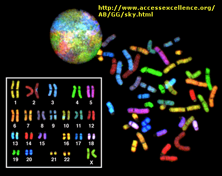
Figure: Human Chromosomes
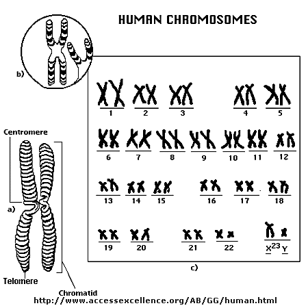
The standard picture of a chromosome with which you are familiar, including the one shown above, is actually
one chromosome of a pair that has just replicated!. One of the chromosomes will stay will the mother cell, and the other will
go to the daughter cell. These two chromosomes which are aligned and appear joined at their centers are called sister chromatids.
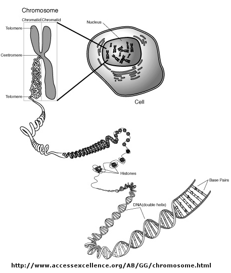
These large DNA/protein complexes must be further packaged in the nucleus, as shown in the "Carl Saganesque" reducing
view of the chromosome, a double stranded DNA molecule winds around a core of proteins.
Figure: Packaging of DNA in the Nucleus
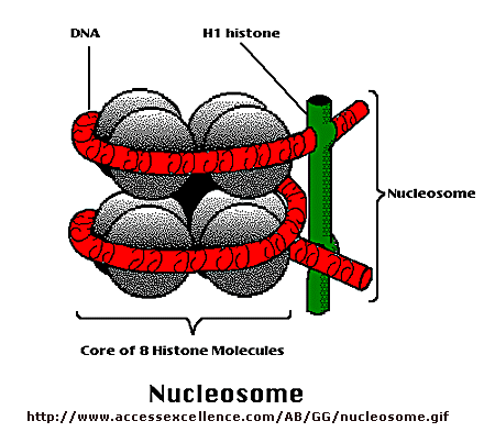
The core is called a nucleosome, and can be viewed under an electron microscope if the chromosome is disperesed.
The nucleosome consists of 8 positively charge proteins called histones. In the core are 2 copies each of His 2A, 2B, 3, and
4. The dsDNA winds around the nucleosome core about 2.5 times. The dsDNA then links around other nucleosome in whch each nucleosome
is connected by a small section of interconnecting spacer DNA, to which is bound another histone, H1. Under an electron microscope,
the DNA looks like a bead on a string. The beads are nucleosomes, and the string is the dsDNA.
Figure: Nucelosome
More DNA Web Links
|
| | | |
|