Polysaccharides
These contain many monosaccharides in glycosidic links, and may contain many branches. They serve as either
structural components or energy storage molecules. The most common polysaccharides consisting of single monosaccharides are:
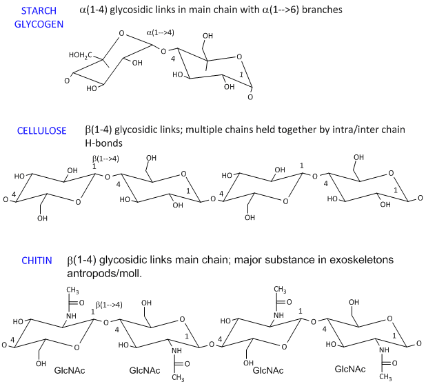
- starch (found in plants). It's a polymer of Glc linked in a main chain through a 1->4 links with a 1->6 branches. Amylose is starch
with no branches, while amylopectin has branches. Starch granules consist of about 20% amylose and 80% amylopectin.
-
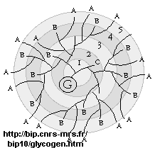
glycogen, the main CHO storage in animals. Muscle and liver glycogen consists of Glc residues
in a 1->4 links with lots of a
1->6 branches (many more branches than in starch). The polymer is synthesized on a protein primer called glycogenin
(G), and has a structure shown below (in which only 5 rings of the structure are shown instead of the actual 12. (Meléndez-Hevia
et al. )
- celluose, a structural Glc polymer in plants, consists of b
1->4 links. It is held together by intra and inter-chain H-bonds. It is the most abundant biological molecule in nature.
- chitin, the major substance in exoskeletons of anthropods and mollusks is a b 1->4 linked polymer of GlcNAc.
The basic chemical structures of these homopolymers are shown below.
Glc b (1-4) Glc link
It makes great chemical sense to store Glc residues as either glycogen or starch, which is one large molecule.
A review of colligative properties would inform you that if all the Glc was stored as the monosaccharide, a great osmotic
pressure difference would be found between the outside and inside of the cell. It makes more sense to have glycogen
exist as a many-branched linear polymer. When Glc is needed, it is cleaved one residue at a time from all the branches (at
the nonreducing ends), producing a large amount of free Glc in a short time.
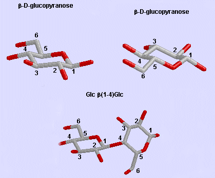
Phi/Psi angles can also be described for the starch/glycogen main chain (around the acetal O) in
a fashion comparable to the for proteins (around the alpha carbon). The phi torsion angle describes rotation around
the C1-O bond of the acetal link, while the psi angle describes rotation around the O-C4 bond of the same acetal link, with
the glucopyranose ring considers as a rigid rotator (just as the 6 atoms in the planar peptide bond unit). The most
extended form of a Glcn polymer occurs when the glycosidic link is B1->4 (as in cellulose), which forms
linear chains. The a 1->4 linked main chain of glycogen and starch
causes the chain to turn and form a large helix, into which can fit iodine (or I3-), which turns starch
purple.
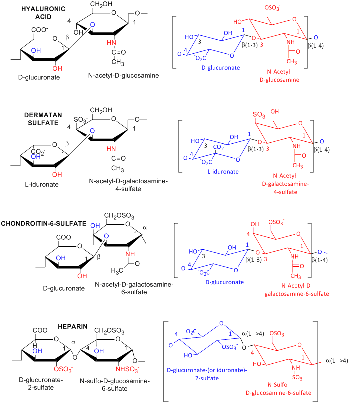
Many polysaccharides consist of repeating dissacharides units. These include the following glycosoaminoglycans
(GAGs), all which contain one amino sugar and in which one or both of the sugars contain negatively charged sulfate or carboxyl
groups. The extent and position of sulfation varies widely between and within GAGs.
- hyaluronic acid, a polymer of Glucuronate (b 1->3)
GlcNAc
- dermatan sulfate, L-iduronate (b 1->3 ) GalNAc-4-sulfate
- keratan sulfate, D-Gal (b 1->4) GlcNAc-6-sulfate
- chondrotin sulfate, D-glucuronate (b 1->3) GalNAc-4
or 6-sulfate
- heparin - D-glucuronate-2-sulfate (a 1->4) GlcNSulfo-6-sulfate
GAGs are found in the vitreous humor of the eye and synovial fluid of joints, and in connective tissue
like tendons, cartilage, etc, as well as skin. They are found in the extracellular matrix and are often covalently attached
to proteins to form proteolglycans.
COMPLEX OLIGOSACCHARIDE DERIVATIVES
1. Cell Membranes of Bacteria
In contrast to eukaryotic cells, bacteria cells have a cell wall in addition to a lipid bilayer membrane.
These are essentially carbohydrate polymers which offer protection from exterior hypotonic condition and the high internal
osmotic pressures, preventing swelling and bursting of the cells. The membrane consist of a peptidoglycan. Two types exists.
a. Gram positive bacteria- These bacterial can be
stained with Gram stain. The wall consists of a GlcNAc (b
1->4) MurNAc repeat (like that in chitin which is a polymer of GlcNAc in (b
1->4) links, but in which the OH of lactate is in ether-linkage to C3 to form N-Acetylmuramic acid). A tetrapeptide
(Ala-D-isoGlu-Lys-D-Ala) is attached in amide link to the carboxyl group of the lactate in MurNAc. The GlcNAc (b 1->4) MurNAc strands are covalently connected by a pentaglycine bridge through the
epsilon amino group of the tetrapeptide Lys on one strand and the D-Ala of a tetrapeptide on another strand.
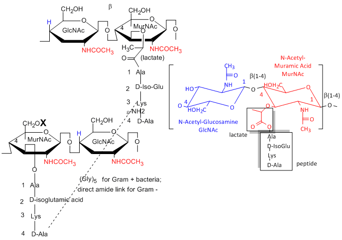
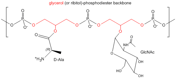
One final structure is found in Gram + membranes. Techioic acids are often attached to the C6 of MurNAc.
Teichoic acid is a polymer of glycerol or ribitol to which alternative GlcNAc and D-Ala are linked to the middle C of the
glycerol. Multiple glycerols are linked through phosphodiester bonds. These teichoic acids often make up 50% of the dry weight
of the cell wall, and present a foreign (or antigenic) surface to infected hosts. These often serve as receptors for viruses
that infect bacteria (called bacteriophages).

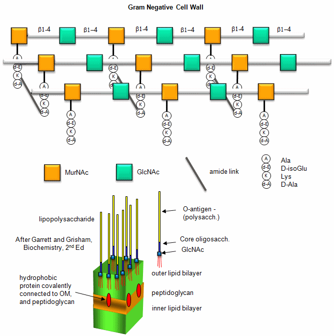
b. Gram negative bacteria-
These bacterial can NOT be stained with Gram stain. The wall consists of the same structure
as in Gram positive bacteria, but the GlcNAc (b 1->4)
MurNAc strands are covalently connected through a direct amide bond between the epsilon amino group of the tetrapeptide Lys
on one strand and the D-Ala of a tetrapeptide on another strand. (i.e. no pentaGly spacer). In addition, Gram negative bacterial
don't have teichoic acid polymers. Rather they have a second, outer lipid bilayer. The cell wall is sandwiched between the
inner and outer bilayers. The space between the lipid bilayers is called the periplasmic space. A hydrophobic protein
covalently attaches (through an amide link from a protein Lys) to the cell wall at the last amino acid in the tetrapeptide
unit of the wall (actually diaminopimelic acid which replaces about 10% of the D-Ala in the cell wall). The N-terminal of
the hydrophobic proteins attaches to the outer lipid membrane through a Ser. The outer membrane is coated with
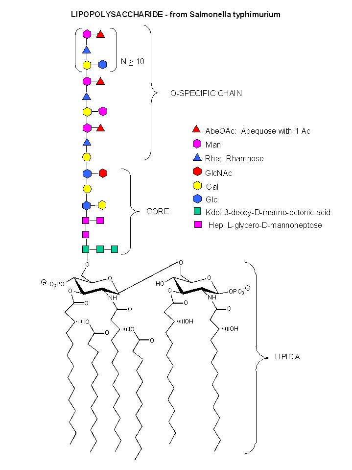
a lipopolysaccharide (LPS) of varying composition. The LPS determines the antigenicity of the bacteria. The different LPS
are called the O-antigens.
Figure: A detailed view of LPS
A recent article (Designing surfaces that kill bacteria on contact by Joerg C. Tiller, Chun-Jen Liao,
Kim Lewis, and Alexander M. Klibanov, Proc. Natl. Acad. Sci. USA, May 22, 2001) describes a new cationic surface which can
kill bacteria on contact. Abstract: "Poly(4-vinyl-N-alkylpyridinium bromide) was covalently attached to glass
slides to create a surface that kills airborne bacteria on contact. The antibacterial properties were assessed by spraying
aqueous suspensions of bacterial cells on the surface, followed by air drying and counting the number of cells remaining viable
(i.e., capable of growing colonies). Amino glass slides were acylated with acryloyl chloride, copolymerized with 4-vinylpyridine,
and N-alkylated with different alkyl bromides (from propyl to hexadecyl). The resultant surfaces, depending on the alkyl group,
were able to kill up to 94 ± 4% of Staphylococcus aureus cells sprayed on them. A surface alternatively created by attaching
poly(4-vinylpyridine) to a glass slide and alkylating it with hexyl bromide killed 94 ± 3% of the deposited S. aureus cells.
On surfaces modified with N-hexylated poly(4-vinylpyridine), the numbers of viable cells of another Gram-positive bacterium,
Staphylococcus epidermidis, as well as of the Gram-negative bacteria Pseudomonas aeruginosa and Escherichia coli, dropped
more than 100-fold compared with the original amino glass. In contrast, the number of viable bacterial cells did not decline
significantly after spraying on such common materials as ceramics, plastics, metals, and wood." This represents a chemical
killing similar to bleach in that a single gene or gene target is not targeted by the surface. The polymer has a permanent
positive charge, which interacts with the negative cell membrane and wall, leading to killing of the cells. A simple
mutation would not make the bacteria resistant to the surface since wholesale changes in gene structure would have to occur
to make tha bacteria resistant.
2. Glycoprotens
Many proteins, especially those destined for secretion or insertion into membranes, are post-translationally
modified by attachment of carbohydrates. They are usually attached through either Asn or Ser side chains. Carbohydrate modifications
on the protein appear to be involved in recognition of other binding molecules, prevention of aggregation during protein folding,
protection from proteolysis, and increases half-life of the proteins. In contrast to a protein sequence which is determined
by the DNA sequence, sugars are attached to proteins by enzymes which recognize appropriate sites on proteins and attach the
sugars. Since there are many sugars which contain many functional groups that can serve as potential attachment sites, the
structures of the oligosaccharides attached to proteins are enormously varied, complex, and hence "information rich" compared
to linear or folded polymers like DNA and proteins.
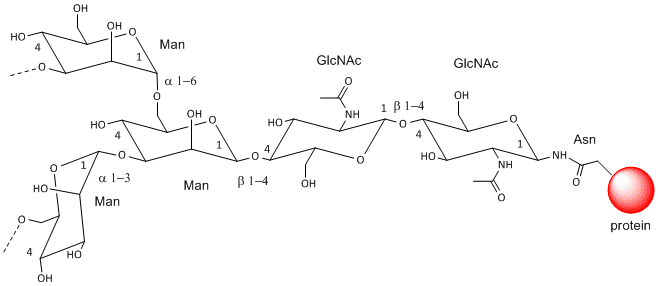
a. N-linked Glycoproteins
These contain CHOs attached through either a GlcNAc or GalNAc to an Asn in a X-Asn-X-Thr sequence of the
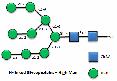
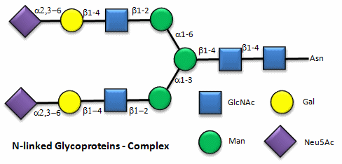
protein. There are three types of N-linked glycoproteins, high mannose, complex, and hybrid. They all contain the same
core oligosaccharide - (Man)3(GlcNAc)2 attached to Asn.
High Mannose N-linked Oligosaccharides
Complex N-linked Oligosaccharides
Hybrid N-linked Oligosaccharides
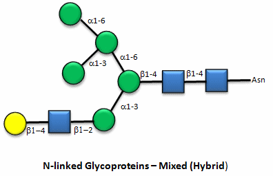
Note that in the hybrid oligosaccharide, one terminus contains Gal(b-1,4)GlcNAc. However, in all other mammals
except man, apes, and old world monkey, an additional Gal is often connected in an a-1,3 link to the Gal to give a terminus of: Gal(a-1,3)Gal(b-1,4)GlcNAc.
These animals have an additional enzyme, a-1,3
Gal transferase. Bacteria also have this enzyme and since we have been exposed to this link through bacterial infection,
we mount an immune response against it. Why is this important? Pig hearts turn out to be similar to human hearts,
so they might be good candidates for transplantation into humans (xenotransplants). However, the Gal-a-1,3-Gal link is recognized as foreign,
and we mount a significant immune response against it. Several biotech firms are trying to delete the pig a-1,3 Gal transferase which would prevent the addition of the
terminal Gal, and make them good donors for transplanted hearts.
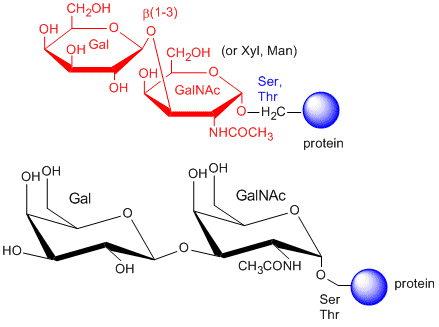
b. O-linked Glycoproteins
The CHOs are usually attached from a Gal (b 1-3) GalNAc
to a Ser or Thr of a protein. The blood group antigens (CHOs on cells attached to either proteins or lipids) are examples
.
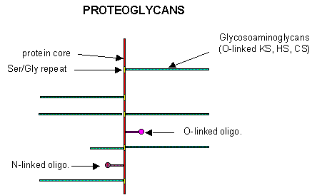
3. Proteoglycans - PGs
Some proteins are so modified with CHOs that they contain more CHOs than amino acids. Proteins linked
to glycosoaminoglycans are together called proteoglycans. (PGs) The structure of a few proteoglycans is known. The GAGs are
O-linked to the protein typically to a Ser of a Ser-Gly dipeptide often repeated in the protein. Some of the proteoglycans
also contained N-linked oligosaccharide groups.
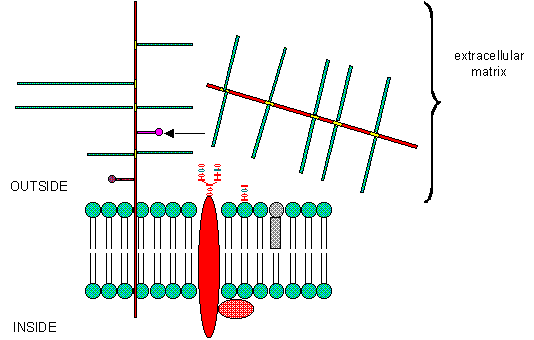
PGs can be soluble and found in the extracellular matrix, or as integral membrane proteins. Given the
diversity of sugars and the varying extent of sulfation, the CHO part of PGs provide an incredible vary of binding structures
to at or near the cell surface. One PG, syndecan, binds through its intracellular domain to the internal cytoskeleton of the
cell, while interacting with another protein - fibronectin - in the extracelluar matirx. Fibronectin also binds other molecules
which can regulate cellular growth and other interactions. PGs act like glue in connecting the extracellular and intracellular
functions of the cell. Most proteins bind PGs through a PG binding motif of BBXB or BBBXXB where B is a basic amino acid.
Some proteins bind to specific sequences in specific GAGs. For instance, antithrombin 3, an inhibitor of blood clotting, binds
specifically to heparin, which enhances its interaction with the clotting protein thrombin.
4. Cell Membranes of Eukaryotes
We have studied lipids, proteins, and carbohydrates. Although phospholipid can spontaneously form biliayers,
the actual structure of biological membranes is made much more complicated through addition of protein and carbohydrate substituents
to the membrane.
We have talked previously about how normally soluble proteins can be made to insert into bilayers by addition
of nonpolar attachments. Several examples of such attachments include:
- N-myristoylation (attached myristic acid - 14:0 - through an amide link)
- S-palmitoylation (attach palmitic acid - 16:0 - through a thioester link with a Cys
- farnesyl or geranylgeranyl additon to a CAAX carboxy-terminal sequence in a target protein, where C is
Cys, A is aliphatic, and X is any amno acid
- addition of a protein to a glycosyl phophatidylinositol (GPI), through a complex
which usually contains a conserved tetrasaccharide core of 3 Man and 1 GlcNAc residues linked to a protein. The GPI can be
further modified with extra Gal's and Man, as well as additions to the PI group, which secures the protein in the membrane.
GPIs are found in eukaryotic cells, and link many surface antigens, adhesion molecules, and hydrolases to the membrane. GPIs
from Plasmoidium falciparum, the malarial parasite which kills about two million people each year, appears to act as a toxin
and is the most common CHO modification of the parasite protein. Mice immunized against the GPI sequence, NH2-CH2-CH2-PO4-Man
(a1-2) 6Man (a1-2) Man (a1-6) Man (a1-4)
GlcNH2 (a1-6) myo-inositol-1,2-cyclic-phosphate, were substantially protected from
malarial symptoms and death after they were exposed to the actual parasite.
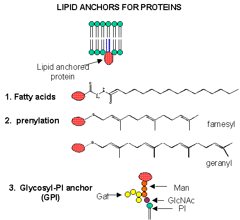
Figure: Biological Membranes: Simple to Complex
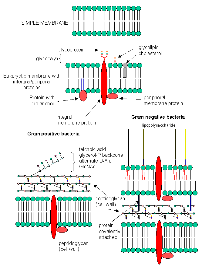
Figure: A cool view of a membrane surface
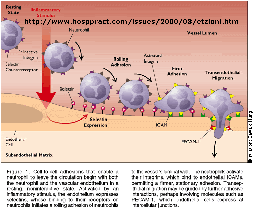
Role of Cell Surface Carbohydrates
Cell surface carbohydrates present information-rich binding sites for other molecules and act as "receptors"
for biological agents as diverse as viruses, bacteria, toxins, and other cells. This is illustrated well by studying
the properties of circulating immune cells. The cells must often pass through the walls of capillaries as they hone
in on a site of infection. (Cancer cells do this as well as they escape the boundaries of the organ in which they developed
and pass through the blood vessels and into new tissues in the process of of forming metastases.) Immune cells must
first bind to endothelial cells (a monolayer of cells that line the lumen of the blood vessels) before they can pass
through the vessel walls. Proteins called selectins our found on cells that can pass through vessels. There are
3 types:
-
L-selectins: found on leukocytes (circulating immune cells)
-
P-selectins: found on activated platelets (which can aggregate to form a type of blood clot)
-
E-selectins: found on activated endothelial cells only after the cells have been induced to form them by certain
immune hormones called cytokines
These selectins are transmembrane proteins with an extracellular CHO binding domain. Such protein domains,
found in all organisms, are called lectins.
Lectins and CHO ligands
| Lectin Family/Lectin |
Abbreviation |
Ligand(s) |
|
Plants |
| Concanavilin A |
ConA |
Mana1-OCH3 |
| Griffonia simplicifolia lectin 4 |
GS4 |
Lewis b (Leb) tetrasaccharide |
| Wheat germ agglutinin |
WGA |
Ner5Ac(a2->3)Gal(b1->4)GlcGlcNAc(b1->4)GlcNAc |
| Ricin |
|
Gal(b1->4)Glc |
|
Animals |
| Galectin-1 |
|
Gal(b1->4)Glc |
| Mannose-binding protein |
MBP-A |
High Mannose Octasaccahride |
| Viral |
| Influenza Virus hemagglutinin |
HA |
Neu5Ac(a2->6)Gal(b1->4)Glc |
| Polyoma virus protein 1 |
VP1 |
Neu5Ac(a2->3)Gal(b1->4)Glc |
|
Bacterial |
| Enterotoxin |
LT |
Gal |
| Cholera toxin |
CT |
GM1 pentasaccharide |
In animals, lectins facilitate cell-cell interactions by forming multiple, but weak interactions between the
protein and many sugars on the ligand to which it binds. The selectins recognize Ser-linked CHO residues (a tetrasaccharide
containing sialic acid, galactose, fucose, and GalNAc) displayed on endothelial transmembrane proteins called
cell adhesion molecules (CAM). These interactions slow the lymphocyte down and activate expression of another protein
on the lymphocyte, integrins. These cause strong lymphocyte-endothelial cell interactions, leading to ultimate
movement of the lymphocytes through the vessel wall. Intergrins are dimeric membrane proteins containing an alpha and
beta subunit.
Genbacev et al. have recently shown that a fertilized egg (in the blastocyst stage which is ready for implantation
in the uterine cell wall) express L-selectin which allows a low affinity (rolling-type) interaction of the fertiized egg with
the uterine epithelial cells. These cells expressed the CHO ligands on their surface which bind to the L-selectin on
the blastocyst. The CHO ligands are only transiently expressed on the surface of the epithelial cells of the uterus,
presumably only when the uterus is primed for implantation. After the initial interaction of the blastocyst and epithelial
cells, further expression of integrins on the blastocyst surface might result. Problems in any of these molecular steps
could result in infertility.
Figure: lectins, integrins, and selectins
 Integrin: The molecular glue of life
Influenza virus binds to sialic acid on host cells, through a binding protein called hemagglutinin. On
binding, conformation, conformational changes activate a neuraminidase activity of a viral protein, allowing cleavage of the
sialic acid glycosidic bond, and subsequent entry of the virus into the cell.
Great CHO Web Sites